Treated by Dr. Roshan Kumar Jaiswal
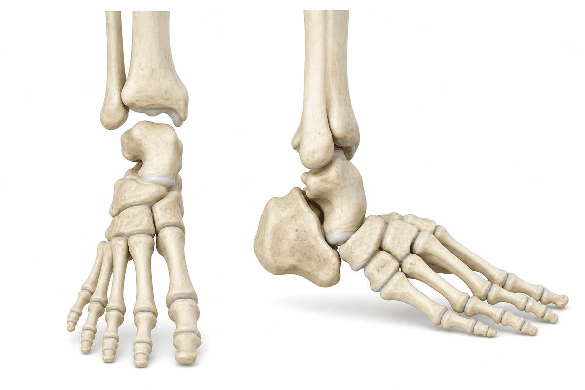
Congenital Vertical Talus is a rare foot deformity present at birth, affecting children in Secunderabad and requiring specialized pediatric orthopedic intervention. The condition causes a rigid flatfoot with a characteristic rocker-bottom appearance, where the sole of the foot is convex rather than having a normal arch. Dr. Roshan Kumar Jaiswal provides expert diagnosis and comprehensive treatment for this complex pediatric foot condition.
Isolated foot deformity occurring without any associated syndromes or neuromuscular conditions, representing the primary form where the vertical talus develops spontaneously during fetal development without identifiable underlying systemic causes.
Vertical talus associated with neuromuscular disorders such as myelomeningocele, arthrogryposis, or genetic syndromes including trisomy 18, requiring comprehensive management of both the foot deformity and underlying systemic condition.
A flexible variant where the foot deformity can be partially corrected manually, demonstrating less rigid contractures and potentially responding better to conservative treatment approaches compared to the rigid forms of the condition.
Multiple factors can contribute to the development and progression of this condition.
Congenital Vertical Talus develops gradually. Recognising symptoms early gives you more treatment options.
From conservative to surgical — we always start with the least invasive option first.
A structured, patient-first approach from first visit to full recovery.
Dr. Roshan Kumar Jaiswal performs a thorough clinical examination of the newborn's feet, assessing flexibility, joint positions, and associated anomalies. Detailed radiographic imaging is obtained to confirm the diagnosis and evaluate the severity of talonavicular dislocation and talus position, while screening for associated neuromuscular or syndromic conditions that may influence treatment planning.
Based on the child's age, deformity severity, and flexibility, Dr. Jaiswal develops a customized treatment protocol. For infants presenting early with flexible deformities, the reverse Ponseti serial casting protocol is initiated. For rigid deformities or older children, surgical intervention is planned with detailed discussion of risks, benefits, and expected outcomes with the family.
When surgery is indicated, Dr. Roshan Kumar Jaiswal performs meticulous soft tissue releases and joint reductions using advanced pediatric orthopedic techniques. The procedure addresses all contracted structures systematically, reduces the talonavicular dislocation, and stabilizes the correction with appropriate fixation. Special attention is given to preserving growth plates and minimizing scarring in growing children.
Dr. Jaiswal implements a comprehensive postoperative protocol including serial cast changes, transition to bracing, and progressive rehabilitation. Regular clinical and radiographic monitoring ensures maintained correction and early detection of any recurrence. Families receive detailed guidance on brace compliance, shoe modifications, and developmental milestone expectations throughout the child's growth.
What to expect at each phase of recovery.
Following surgical correction, the foot is immobilized in a long leg cast for 8-12 weeks to allow soft tissue healing and maintain joint reductions. Pin removal is performed at 6-8 weeks, typically in clinic without anesthesia. Parents are educated on cast care, circulation monitoring, and signs of complications requiring immediate attention.
After cast removal, the child transitions to custom ankle-foot orthoses (AFOs) or specialized bracing to protect the correction and prevent recurrence. Initial full-time brace wear gradually transitions to nighttime-only use based on the child's age and correction stability. Physical therapy focuses on gentle range of motion, strengthening, and age-appropriate motor development activities.
Regular follow-up visits every 3-6 months monitor foot position, growth, and function throughout childhood. Radiographic surveillance detects early signs of recurrence requiring intervention. Brace modifications accommodate growth, and eventual transition to supportive footwear occurs when skeletal maturity and correction stability are confirmed. Gait analysis and functional assessments guide any additional interventions needed.
With appropriate treatment initiated in infancy, most children achieve plantigrade foot position with normal heel contact during walking. The rocker-bottom deformity is corrected, allowing proper weight distribution and improved biomechanics. Children can wear regular shoes and participate in age-appropriate activities without significant limitations.
Correction of the vertical talus deformity enables development of more normal walking patterns and reduces compensatory abnormalities at the ankle and knee. While some residual stiffness may persist, functional mobility is significantly enhanced compared to untreated deformity. Most children achieve independent ambulation without assistive devices.
Proper foot alignment eliminates abnormal pressure points that cause painful callosities on the plantar prominence. Normalized weight-bearing patterns prevent chronic pain associated with walking on the midfoot. Long-term comfort is significantly improved, supporting active participation in daily activities and sports.
Early intervention prevents progressive compensatory changes in the ankle, knee, and hip joints that develop with untreated vertical talus. Addressing the deformity during infancy takes advantage of skeletal plasticity, minimizing the need for extensive reconstructive procedures later. Risk of degenerative arthritis in adulthood is reduced through anatomic restoration.
Untreated congenital vertical talus leads to lifelong disability with severe pain, difficulty walking, and inability to wear normal footwear. The rigid rocker-bottom foot causes abnormal gait patterns and progressive secondary deformities in the ankle, knee, and hip joints. Chronic pain from callus formation on the plantar prominence, limited mobility, and significant functional impairment severely impact quality of life and may require more complex reconstructive surgery in adolescence or adulthood with less favorable outcomes.
Parents should seek immediate evaluation by a pediatric orthopedic specialist if they notice an abnormal rocker-bottom foot shape in their newborn or infant, particularly if the sole appears convex rather than having a normal arch. Early diagnosis and treatment initiation, ideally within the first few months of life, significantly improve outcomes and may allow for successful conservative management. Any child with known neuromuscular conditions or genetic syndromes should receive orthopedic screening for associated foot deformities including congenital vertical talus.
Early treatment means more options and better outcomes. Book a consultation to understand your condition and explore the right path forward.
Consult Roshan Kumar Jaiswal for Congenital Vertical Talus at any of these 9 centres — pick the one nearest you.